|
|
Welcome & Introduction | |
Basics & Anatomy | |
Treatment of a Case | |
Resources & References | |
Clinical Trials | |
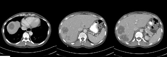
Case: 37 RADIOLOGIC ASSESSMENTDiscussant: FERGUS COAKLEY, MD (Dr Alberts Case)Images:These four contrast-enhanced CT images were obtained in the portal venous phase. Clearly, two large metastases are seen in the right hepatic lobe, in segments VII and VIII, superiorly. Turning to the left lobe, we can identify on that second image an approximate 1.5-cm metastasis on the posterior aspect of the left lateral segment. On image number three, a possible small lesion is suggested — I’m not absolutely certain — in the anterior aspect of the left lobe. Finally, on the fourth image, we see somewhat eccentric, cystic-appearing wall thickening of the descending colon — which is presumably the known primary tumor — possibly with some areas of necrosis or extramural extension, perhaps even a “so-pre-ent” section with an abscess formation. All of those things would be possible radiologically. The second image shows what appears to be the middle hepatic vein. A reasonable margin seems to be present between that and the metastasis in segment VIII. When evaluating vessels, it is helpful to have as many images as possible.
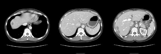
Postchemotherapy scan:Three contrast-enhanced CT images were obtained in the portal venous phase, showing a dramatic response to chemotherapy, with marked shrinkage of the two metastases in the right hepatic lobe. I can no longer see the two areas of concern in the left lobe, so this patient has biologically shown a good response to chemotherapy and would seem to be a good candidate for surgery. Of course, one of the downsides of performing surgery after chemotherapy is that you never know for sure if you’ve missed your chance to conduct the best possible staging of the peritoneal cavity and the nodes.
|
|