|
|
Welcome & Introduction | |
Basics & Anatomy | |
Treatment of a Case | |
Resources & References | |
Clinical Trials | |
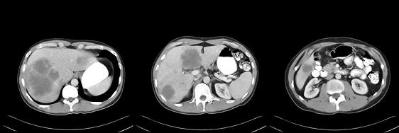
Case: 38 RADIOLOGIC ASSESSMENTDiscussant: FERGUS COAKLEY, MD (Dr Alberts Case)The initial abdominal CAT scan: Three contrast-enhanced CT images were obtained in the portal venous phase. We see several large hepatic metastases. One is occupying almost the entire upper right lobe. One is in the medial segment of the left lobe, one is on the lateral segment of the left lobe and one is in the inferior right lobe, so certainly on this baseline study, it’s hard to conceive of any surgery that could remove all of these lesions and still leave the patient with a reasonable amount of residual liver function.
Postchemotherapy:We can see a dramatic response of the disease, with many of these lesions shrinking to the point of invisibility or being much smaller, and appearing quite bland and nonspecific on the postchemotherapy scans. This speaks to the need to examine the previous CT images as your baseline, because when viewing the postchemotherapy images alone, you might not even know that these were metastases.
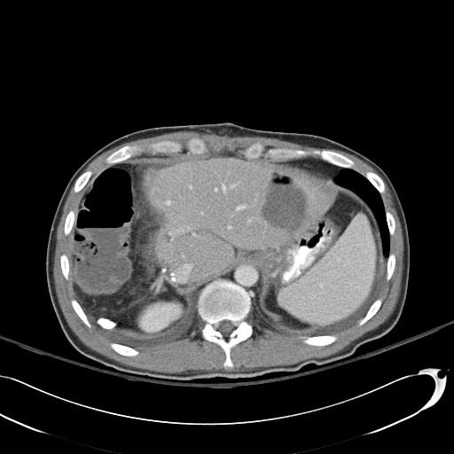
Postoperative scan, day six:This is a contrast-enhanced CT image showing changes related to a right hepatectomy, and then also an air- and fluid-containing lesion on the left lobe. As a radiologist, it is important to realize that this could be entirely within the normal postoperative range of appearances for an ablation site, be it a radiofrequency ablation (RFA) or wedge resection. They can certainly have both air and fluid in the ablation site. The importance is not to get overly excited or call this an abscess necessarily or suggest that it indicates infection. This is within the range of expected postoperative appearances.
|
|