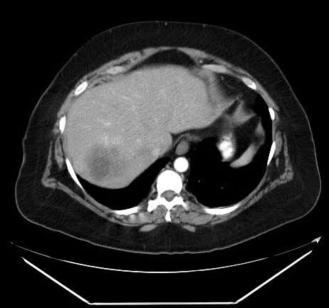
A somewhat heterogeneous, low-density mass is shown in the right lobe involving segment VII and VIII of the liver. This is a relatively arterial phase image, so the aorta is bright. This is not the best phase of contrast enhancement to evaluate the liver for suspected colorectal metastases. The use portal phase imaging is preferred.
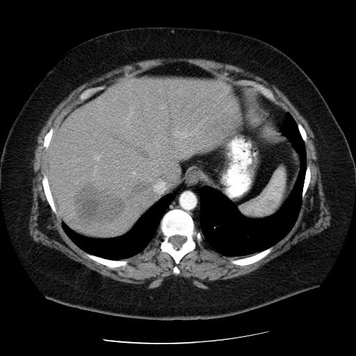
Post-neoadjuvant image: 6/08
The low-density lesion looks larger. Although it could be argued that the edges are improved, this image was taken in the arterial phase. Although not common, multiphasic studies — arterial phase, portal phase, delayed phases in all of their protocols — are sometimes used.
As a general principle, people think that if they observe lower density, blacker material than on prior study, it signifies necrosis. Although it could be necrosis, it could also be attributed to the variable timing of the studies. For example, this case includes an arterial phase image. If you compared this image to one from a portal phase, this (the lesion?) may begin to get whiter and pick up contrast.
It is extremely dangerous to talk about necrosis using one snapshot image — particularly in this case, in which you’ve taken the image so early in the time course that you may not even be enhancing anything. You may not have any enhancement in here, because the image was taken so early in this particular phase.