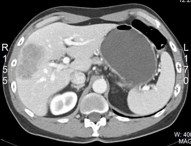
A large lesion, approximately six to seven centimeters in size, is seen in the right lobe of the liver. The lesion has the classical features of a metastasis. This appears to be a segment-VIII lesion. The lesion appears to be free of vessel involvement, according to one cut. Still, one has to be careful. If we had the whole image set, we would be able to track the right main portal vein to see how close the lesion is on the higher cuts.
Follow-up scan:
The lesion now appears to be about five centimeters, indicating a nice response to chemotherapy. On this image, there appears to be separation from the portal venous structures.