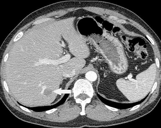
The contrast-enhanced CT image shows an approximate 2- to 3-cm suspicious lesion in segment VII. The left lobe appears relatively small.
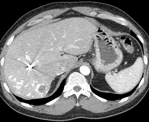
Postembolization image:
The postembolization CT shows the lesion, and the streak artifact from the metallic coils that have been placed in the right portal vein. The left lobe is now significantly larger than it was before.
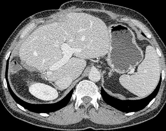
Postsurgical image:
The metastasis has been resected and further hypertrophy of the residual liver has occurred, which shows good volume on this study.