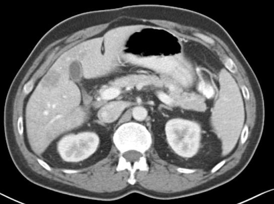
This lesion is approximately four to five centimeters in size with a 2-cm lesion tucked away in the right lobe. Another lesion appears in the interior aspect of the liver. The two- to three-cm lesion is in segment V.
One lesion appears to be abutting right on the right main portal vein. Another lesion appears to be located halfway between segment VIII and IV.
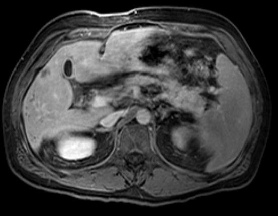
Post-embolization image:
This MRI image shows a lesion traversing the segment VIII and VII. Although it is difficult to make a direct comparison with the prior imaging study — because we are comparing MRI results with those of a CT scan — the lesion appears to be smaller.