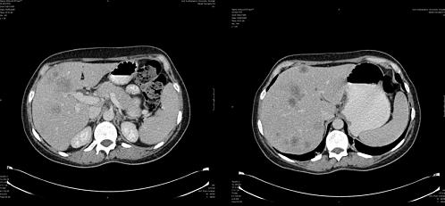
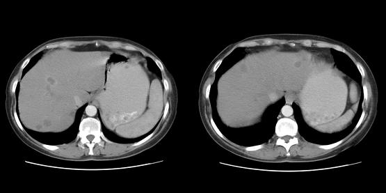
This liver CT shows bilateral low-density lesions — most likely in segment II — and multiple lesions in the right lobe. The bilobar liver lesions are entirely consistent with the history of metastatic disease.
The lesion in segment IV, close to the portal vein, is probably four to five centimeters in size. The lesions vary in size, with the smallest being approximately one centimeter. The lesions occupy almost every segment of the liver.
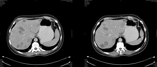
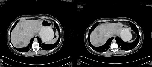
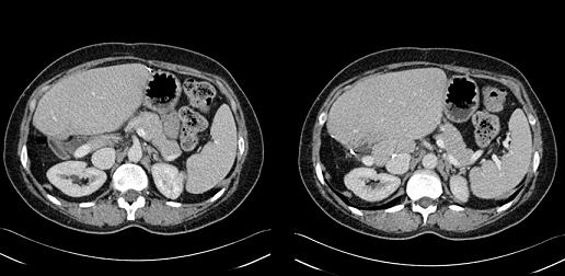
Follow-up CT after chemo/biologic therapy:
This image is a somewhat arterial phase image, which isn’t ideal. The scan suggests that the patient had a nice response to therapy. However, it looks as though a lesion remains in the left lobe, probably in segment II, and on the images provided, it looks as though a lesion is encroaching into segment IV.
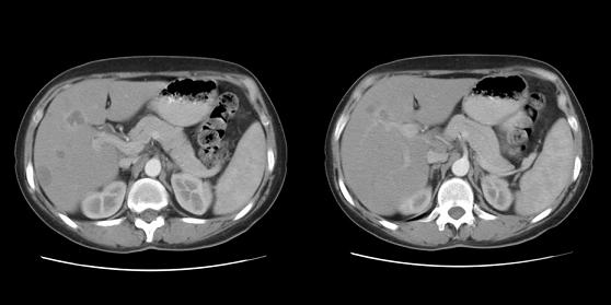
Postoperative image:
This image shows that the resection margin and the liver are now hypertrophied. A recurrent mass or residual mass may be present in segment IV. It could be a site of post ablation or simply residual postoperative change along the resection margin.