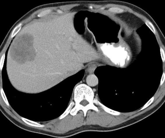
This is a contrast-enhanced CT image showing an approximate 4-cm suspicious hypodense mass in the superior aspect of the liver, probably involving segment VII and VIII.
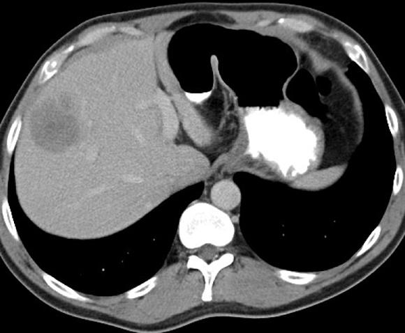
The next image, at a slightly more than inferior level, shows that similar suspicious mass in segment VIII.
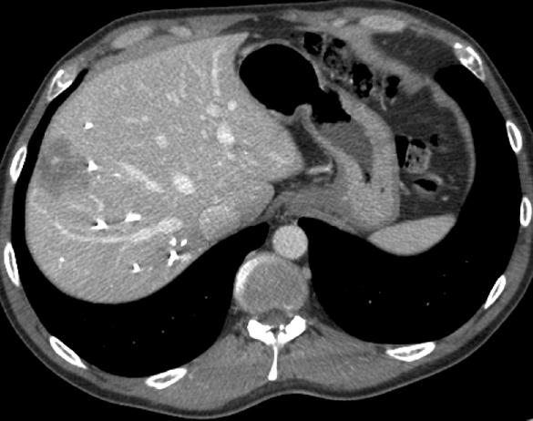
Finally, the lowest image again shows that mass in segment VIII at the level of the porta hepatis. The left lobe is quite small — only a relatively diminutive amount of liver is visible to the left of the portal vein.
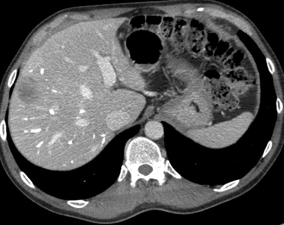
Post-treatment images:
The patient now has had portal vein embolization (PVE) and chemotherapy. We can see that the two hepatic metastases have shrunk considerably. We see a high density of embolic material in the branches of the right portal vein. In particular, on the final image taken on the level of the portal vein, we can appreciate that some hypertrophy has occurred of the previously quite small left hepatic lobe.
Liver Image:
The contrast-enhanced image of the liver obtained in the portal venous phase shows two centrally located hepatic metastases in segment IV that are touching, and in the right lobe, centrally, involving multiple segments of the right lobe. These are adjacent to the portal vein and the portal vein bifurcation, so they’re in a critical anatomic location.
Postchemotherapy:
A good response is observable in the liver metastases. They have shrunk down nicely, but it is still going to be challenging to treat this patient. The posterior lesion will come out with a right lobectomy, but that still leaves the lesion in segment IV — that presumably could also be ablated or perhaps wedged out, in order to render this patient tumor-free.
Postsurgical image:
The patient had a right hepatectomy and the lesion in segment IV, where fluid has collected, may have been treated with a wedge resection. In passing, I would say that these fluid collections adjacent to the resection margin posthepatectomy are extremely common. Most of the time, they’re sterile bilomas, and therefore, they don’t need intervention. Occasionally, if patients are septic or if the fluid collections are extremely large, they may need drainage. The residual liver has experienced satisfactory hypertrophy. We can see how much larger the left lateral segment in particular has become, which is, of course, one of the great features of the liver — how well hypertrophy occurs postresection.