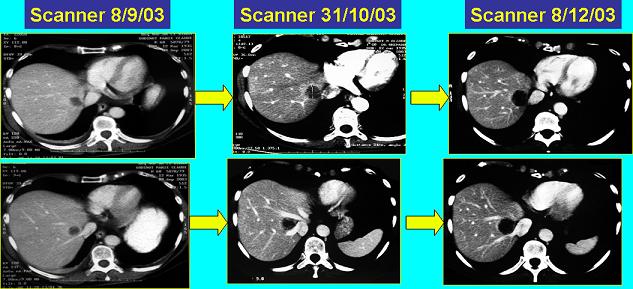
On the initial study, we identified a low-density lesion, probably about 1.5 centimeters, lying anterior to the right hepatic vein and near the cava, so it is in the medial aspect of segment VII.
Postcolectomy scan and preoperative postchemotherapy scan:
On the bottom image, the lesion is relatively well circumscribed, where on the upper image (the baseline), we can see it’s also a bit irregular, with the suggestion of some surrounding hyperemia or increased density. I would be suspicious that this was a metastasis. Certainly we see that it grows over the serial studies, so clearly is metastatic on the follow-up studies.
Perhaps the center of this lesion has more low density, which could suggest some central necrosis. But the overall size of the lesion is probably larger than before. The lesion clearly is abutting both the middle and right hepatic vein, and it’s centrally located near the cava — it is clearly in a critical location with respect to the blood vessels.