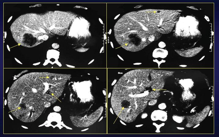
These are four contrast-enhanced CT images showing at least one large suspicious lesion in the right hepatic lobe in segment VII, and then four smaller lesions in the left lobe that are less specific, but certainly concerning for additional sites of disease. The large right-lobe lesion is close to some hepatic vein branches, but I think would not interfere with performing a right hepatectomy.
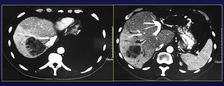
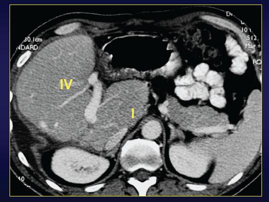
Postchemotherapy scan:
There are four contrast-enhanced CT images post-treatment. The right-lobe lesion that had been the dominant lesion before, appears somewhat larger — certainly, I don’t see any evidence of response. In addition, approximately three additional small lesions are present in the left lobe that appear relatively stable. That may be additional sites of disease, and it seems to be confined to the lateral segment.