MetResect
An educational tool to assist in the management of hepatic metastases in patients with colorectal cancer.

Welcome Methods Authorship Strengths/Weaknesses
Using MetResect Principles Anatomy Tutorial
Selection Options By Category By Faculty By Case Matching
Overview of Resources References by Author References by Topic Expert Comments by Topic
North American Asian European World Wide


Case 25: RADIOLOGIC ASSESSMENT

      Film Review  

Discussant: FERGUS COAKLEY, MD (Dr Adam Case)

CT scan before colectomy:

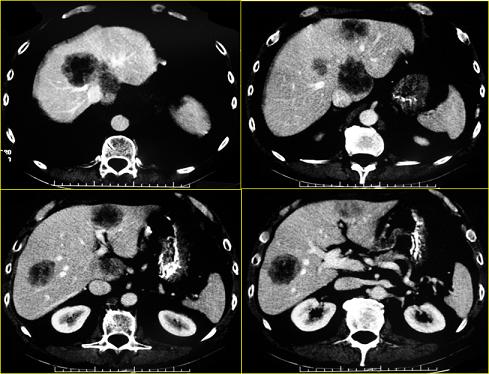
These are four contrast-enhanced CT images obtained in the portal venous phase, showing several hepatic metastases. Of note, both the left and right lobes appear to be involved, with a metastasis in the caudate lobe, which is technically challenging to resect. The lesions in segment IV and the caudate lobe are partially confluent and sitting right at the hepatic venous confluence with the inferior vena cava. In addition to multiplicity involving segments that are difficult to resect, a vascular contact issue is also evident that will make these lesions extremely challenging to resect from a surgical perspective.


Copyright © 2008-2009 Research To Practice. All Rights Reserved.

Contact/Evaluate MetResect