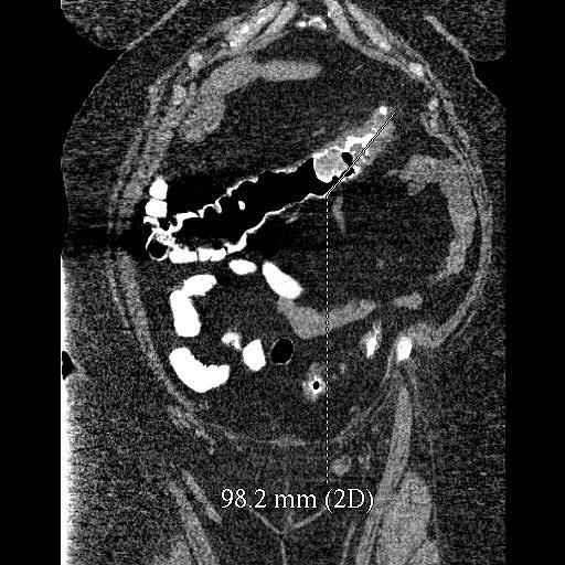
The image shows a nodular thickening in the wall of the distal transverse colon. The white material is barium. The inference is that we have a mucosal lesion that is causing stricturing of the lumen, and this is entirely consistent with a colonic primary carcinoma.
The quality is somewhat degraded by this barium — I don’t understand why barium was used in this study.
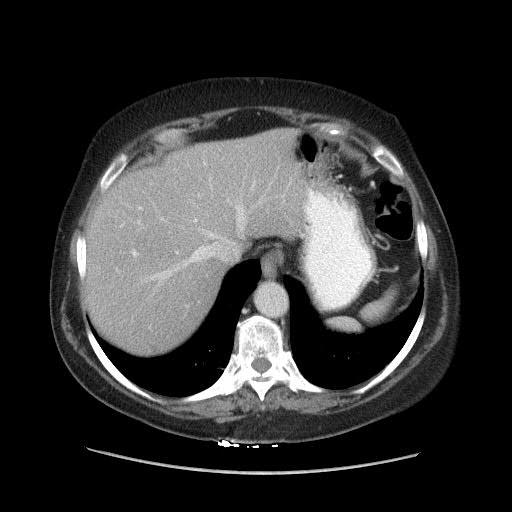
This portal phase study shows a total of three lesions. One lesion in the right lobe of the liver is located peripherally, nowhere near vessels. It is probably about two centimeters in size and is located in segment VII. Another lesion, approximately one centimeter, is in segment VIII.
This is a somewhat fuzzy, grainy image, so it is rather difficult to be sure if other lesions are present elsewhere. One would have to be careful about interpreting these findings, if this was the only cross-sectional image available for this patient.
You see a lot of artifacts from these lines coming across the image, with gray lines everywhere. This particular image is compromised by the CT quality. If a lesion is present, it is located at a bad position, right at the confluence of the right hepatic vein, middle hepatic vein and IVC.