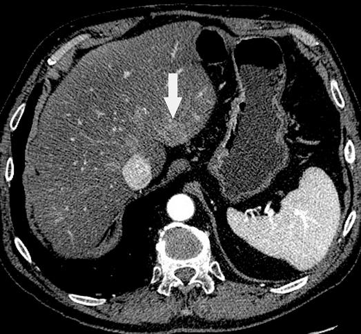
The contrast-enhanced CT image, obtained in the portal venous phase, shows a single, subtle, approximate 3-cm lesion in segment II posteriorly. The liver generally appears to be low density and is likely affected by diffuse fatty infiltration, which we know can limit our sensitivity for metastasis. It is not known whether that fatty infiltration was present at baseline or if it developed as a consequence of chemotherapy. This is a situation where PET can be quite helpful.
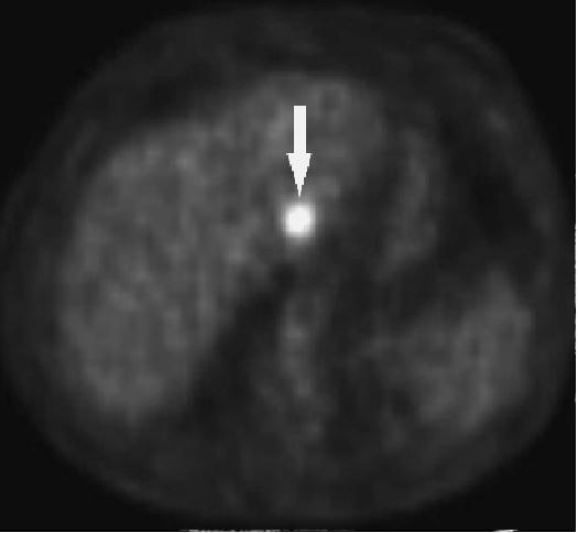
On the PET image, there is absolutely no doubt that there is a focus on increased uptake, corresponding to the subtle lesion seen on CT, confirming that it is indeed metastasis