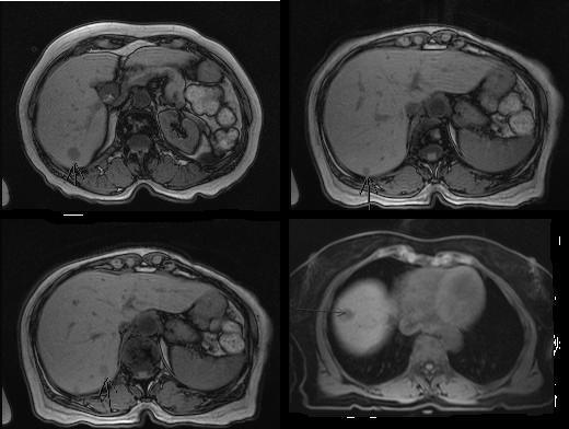
These are noncontrast MRIs. This may be because the patient was not able to receive contrast for some reason or other. A 1- to 2-cm lesion is seen in the periphery of the right lobe, probably in segment VII or VI. In segment VII, there appears to be a sub-centimeter lesion — approximately seven to eight millimeters. Up in the dome of the liver in segment VIII, another lesion is present, probably 1 to 1.5 centimeters in size.
If these noncontrast images were presented without a patient history, they look like what we would call T1-weighted images — I could not tell you what the images were. They could be cysts, hemangiomas or metastases. Additional sequences would be required to confirm the presence of metastasis.
Even if the images were more diffuse and not as circumscribed as they are, I still could not confirm whether or not the lesions were metastatic because they could be abscesses. Like metastasis, abscesses also have a propensity to be ill-defined and have fuzzy margins. You need the clinical history to help you. Based on the imaging alone and this set of images, we wouldn’t be able to tell you what this was. But we know that this person did have liver metastases from other imaging.