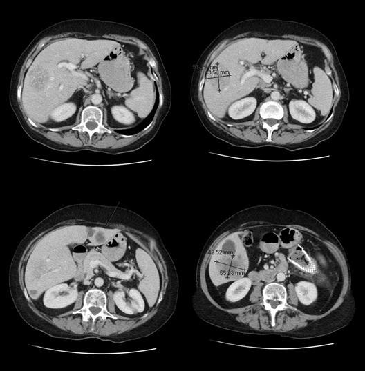
A 4-cm lesion is shown that straddles across several segments in the right lobe, out toward the periphery — it is away from the right branch of the portal vein. A couple of other small lesions are also present. In the left lobe, there is a two- to three-cm lesion that appears to be in segment III of the liver, in the lateral left lobe. Another lesion seems to be straddling the dividing line that goes through the fissure.
The cut above:
This cut shows the line that would divide the lateral left lobe, segments II and III, from the right lobe. The lesion looks to be sitting right on it with another lesion in what looks like segment III. So the disease looks bilobar in the left and right lobe, although in these images, only one lesion is seen in the lateral left lobe and another one seems to be possibly straddling into segment IV.
Last image:
This image shows a metallic stent that was probably put through the obstructing lesion in the transverse colon.