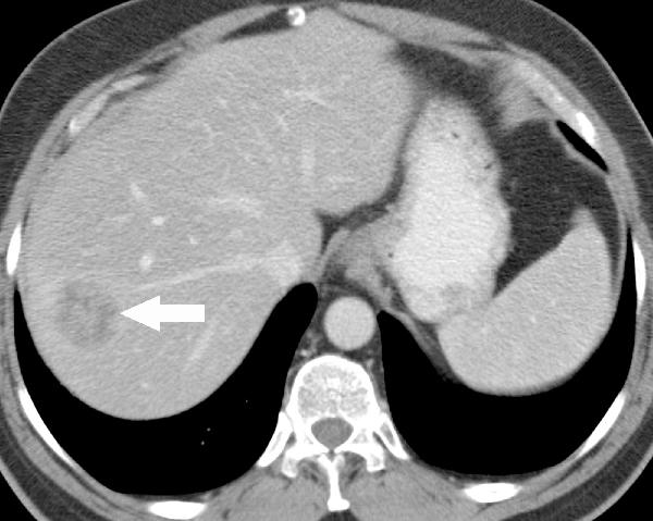
This is a contrast-enhanced CT image through the upper part of the liver showing an approximate 4-cm hypodense suspicious mass in the lateral aspect of segment VII.
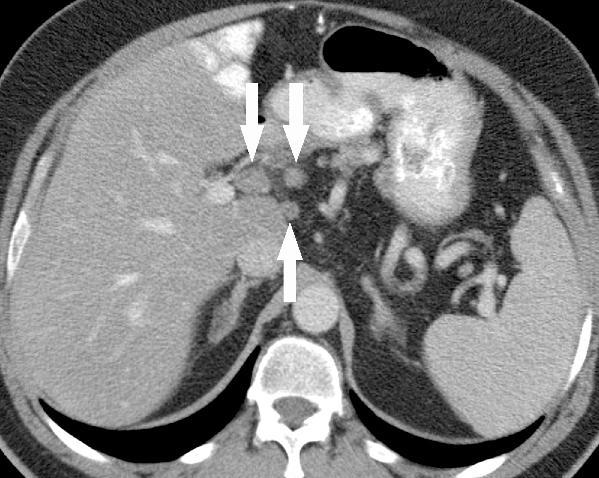
CT image of the porta hepatis:
This image shows three enlarged porta hepatis nodes as indicated by the arrows. The nodes have a kind of rounded appearance, measure up to approximately two centimeters and are at least relatively suspicious for metastasis by CT.
PET scans:
This is a single image from a contrast-enhanced CT showing a large, probably 5-cm suspicious hypodense mass in segment VII, abutting both the IVC and the right hepatic vein. This is in a somewhat difficult location surgically.