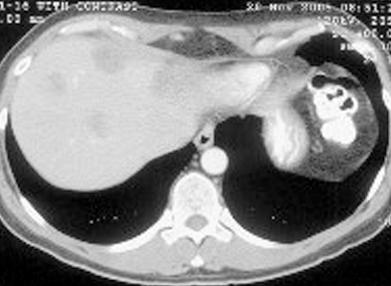
This is a contrast-enhanced CT image obtained in the portal venous phase through the upper liver. Several low-density, suspicious hepatic masses are visible, involving both the visible portion of the left lobe, likely in segment IV and also involving the right hepatic lobe.
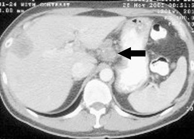
The next CT image again shows the suspicious mass in the right hepatic lobe in segment VIII and then in addition, we see a relatively large porta hepatis nodal mass, highly suggestive of metastatic disease.
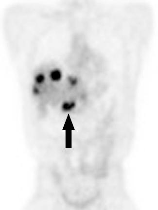
The final image is a PET scan showing unequivocal uptake within the porta hepatis mass, confirming its malignant nature. In addition, we can see several increased foci of uptake within the liver, again confirming bilobar hepatic metastasis and, of note, no additional disease sites are visible on this PET image.